The human skeletal system is an intricate structure that supports the body, allows movement, and protects vital organs. Among the many bones that make up this system are the phalanges, which form the fingers and toes. When studying anatomy or reading medical descriptions, one may encounter the term distal phalanx. This raises a common question is the distal phalanx a bone, or is it something else? Understanding this term requires exploring the anatomy of the hand and foot, the role of the phalanges, and how they contribute to daily movement and fine motor control.
Understanding the Structure of the Phalanges
The word phalanx refers to the bones that make up the fingers and toes. In both the hands and feet, the phalanges are divided into three sections the proximal phalanx, the middle phalanx, and the distal phalanx. Each of these bones plays a specific role in movement and stability. The distal phalanx is the outermost segment, located at the tip of each finger and toe. Therefore, yes, the distal phalanx is indeed a bone – the last bone at the end of each digit.
In the human hand, there are fourteen phalanges in total three for each finger and two for the thumb. The thumb only has a proximal and distal phalanx, which is why it is shorter and more flexible. Similarly, in the foot, each toe has three phalanges except for the big toe, which has only two. The distal phalanx forms the fingertip or the end of the toe, giving shape and structure to the nails and supporting soft tissues.
Location and Function of the Distal Phalanx
The distal phalanx is located at the tip of each digit. In the hand, it supports the fleshy pad that allows for precise manipulation and touch sensitivity. In the feet, the distal phalanges play a crucial role in balance, stability, and propulsion during walking or running.
One of the main functions of the distal phalanx is to provide an anchor for the nail bed. The bone helps shape the fingertip, giving the nail a firm surface to attach to. It also supports the pulp of the finger, the soft tissue that allows people to grip, type, write, and perform other delicate tasks. The distal phalanx is vital for fine motor skills, making it an essential component of the hand’s anatomy.
Structure and Shape
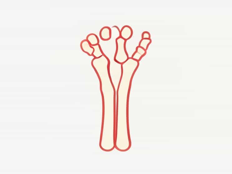
The shape of the distal phalanx is slightly flattened and wide at the end, providing a broad base for the nail and fingertip tissues. The bone narrows toward its proximal end, where it articulates with the middle phalanx (or the proximal phalanx in the case of the thumb and big toe). The distal phalanx contains bone marrow and is covered by a thin layer of periosteum, a membrane that supports bone growth and repair.
Comparison Between Distal, Middle, and Proximal Phalanges
Although all phalanges share a similar basic structure, there are subtle differences between the distal, middle, and proximal segments. The proximal phalanges are the longest, forming the base of the fingers and toes. The middle phalanges act as connectors, and the distal phalanges form the tips.
- Proximal phalanxConnects directly to the metacarpal or metatarsal bone, providing the main support for finger or toe movement.
- Middle phalanxActs as a hinge between the proximal and distal segments, aiding flexibility and motion.
- Distal phalanxProvides structure to the fingertip or toe tip, supporting tactile function and protection.
The distal phalanx differs in size and shape across different fingers and toes. For example, the distal phalanx of the thumb and big toe is broader and stronger, adapted for gripping and weight-bearing.
Bone Composition and Strength
Like other bones, the distal phalanx is composed of compact bone on the outside and spongy bone inside. The compact bone provides strength and rigidity, while the spongy bone contains red marrow that produces blood cells. Despite being small, the distal phalanx is remarkably strong and can withstand everyday use. However, because it is located at the fingertip, it is also vulnerable to injuries such as fractures, crush injuries, or nail bed trauma.
Common Distal Phalanx Injuries
Since the distal phalanx is exposed and used frequently, it is prone to certain injuries. The most common include
- FracturesOften occur from crushing injuries, such as slamming a finger in a door or dropping a heavy object on a toe.
- Mallet fingerHappens when the tendon that straightens the finger is damaged, often accompanied by a distal phalanx fracture.
- Nail bed injuriesThe distal phalanx supports the nail matrix, and damage to this bone can affect nail growth or appearance.
These injuries may seem minor but can significantly affect daily activities if not properly treated. Treatment typically includes splinting, rest, and in some cases, surgical repair.
Development and Growth of the Distal Phalanx
The distal phalanx begins to develop during fetal growth, forming from cartilage that gradually ossifies into bone. Each phalanx has a growth plate (epiphyseal plate) that allows the bone to lengthen during childhood and adolescence. Once a person reaches skeletal maturity, usually in their late teens or early twenties, these growth plates close, and the bones stop growing in length.
This process ensures that the distal phalanges grow proportionally with the rest of the hand and foot, maintaining balance and coordination. The tips of the fingers are among the last areas to fully ossify, reflecting the importance of precise development for fine motor control.
Evolutionary Perspective
From an evolutionary standpoint, the distal phalanges have played a crucial role in the development of human dexterity. The broad and flattened shape of the distal phalanx in humans allows for the attachment of nails rather than claws, supporting precise and delicate movements. This adaptation distinguishes humans from many other primates and is key to tool use, writing, and manipulation of small objects.
Clinical Importance of the Distal Phalanx
In clinical settings, the distal phalanx serves as an important reference point in diagnosing hand or foot conditions. Radiographs (X-rays) often reveal the state of this bone, showing signs of fractures, infections, or degenerative diseases. In addition, the distal phalanx can be affected by conditions such as osteomyelitis (bone infection), psoriatic arthritis, and peripheral vascular disease.
Surgeons and medical professionals must understand the anatomy of the distal phalanx when performing procedures like nail bed repair, fingertip reconstruction, or amputation. The bone’s close relationship with nerves, blood vessels, and soft tissue makes precise handling essential.
In summary, the distal phalanx is indeed a bone – the small yet vital structure located at the tip of each finger and toe. It provides shape, support, and function to the fingertips and toes, allowing humans to perform precise tasks and maintain balance. From a structural, functional, and evolutionary perspective, the distal phalanx demonstrates how even the smallest bones play a major role in human anatomy. Understanding its location, purpose, and importance helps in appreciating the complexity and elegance of the skeletal system as a whole.