Non-ossifying fibroma is a benign bone lesion commonly found in children and adolescents, often discovered incidentally during imaging studies such as knee X-rays. These lesions typically develop in the metaphysis of long bones, particularly around the knee, including the distal femur and proximal tibia. Most patients are asymptomatic, and the condition is often identified when an X-ray is taken due to minor trauma or unrelated issues. Despite being benign, understanding the features of non-ossifying fibroma on knee X-ray is important for differentiating it from other bone lesions and ensuring proper monitoring or treatment if necessary.
Understanding Non-Ossifying Fibroma
Non-ossifying fibroma (NOF) is classified as a fibrous cortical defect that is composed of fibroblasts, histiocytes, and occasional giant cells. It is the most common benign bone lesion in children and adolescents, with a prevalence of up to 30% in individuals under 20 years old. Although NOFs are generally self-limiting and resolve spontaneously over time, they are clinically significant when they are large or located in weight-bearing bones, as they can occasionally weaken the bone and increase the risk of fracture.
Common Locations Around the Knee
The knee is one of the most frequent sites for non-ossifying fibroma. Within the knee region, NOFs typically occur in
- Distal FemurAlong the metaphyseal region just above the knee joint.
- Proximal TibiaBelow the knee joint in the upper portion of the tibia.
- FibulaLess commonly, lesions may be present near the head of the fibula.
The metaphyseal location is key to diagnosing NOF, as lesions in this area usually have characteristic radiographic features that help distinguish them from more aggressive bone tumors.
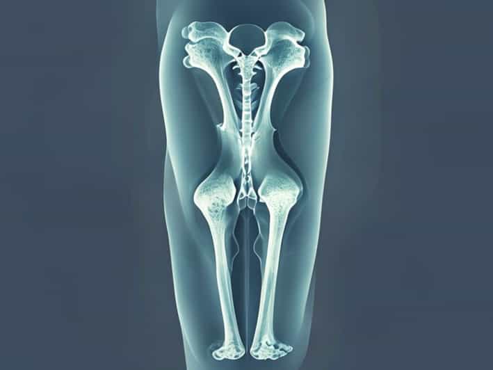
Knee X-Ray Features of Non-Ossifying Fibroma
When examining a knee X-ray for a potential non-ossifying fibroma, several radiographic features are typically noted. Recognizing these features helps radiologists and orthopedic specialists differentiate NOFs from other bone lesions and avoid unnecessary interventions.
Characteristic X-Ray Appearance
Non-ossifying fibromas on knee X-ray generally display
- Well-Defined MarginsThe lesion often has a sclerotic border, which indicates a slow-growing benign process.
- Ovoid or Eccentric ShapeMost NOFs appear as oval or elongated defects in the metaphysis, usually off-center within the bone cortex.
- Radiolucent CenterThe interior of the lesion appears less dense on X-ray due to its fibrous content.
- Thin Cortical RimA thin layer of bone may surround the lesion, maintaining structural integrity while clearly outlining the defect.
- Absence of Periosteal ReactionIn uncomplicated cases, there is typically no periosteal reaction, distinguishing NOF from aggressive bone tumors.
These features help reassure clinicians and patients that the lesion is benign and often requires only observation rather than immediate intervention.
Size and Risk Assessment
The size of the NOF observed on knee X-ray is an important factor. Small lesions are typically asymptomatic and rarely require treatment. Larger lesions, particularly those occupying more than 50% of the bone diameter, may weaken the bone and pose a fracture risk. In such cases, monitoring through periodic X-rays is recommended, and surgical intervention may be considered if fractures or structural compromise occur.
Symptoms and Clinical Presentation
Many non-ossifying fibromas are discovered incidentally because they rarely cause symptoms. However, some patients may present with
- Mild localized pain around the knee
- Swelling or tenderness in the affected area
- Pathological fractures following minor trauma if the lesion is large
Physical examination may reveal localized tenderness, but most children and adolescents with NOFs do not exhibit significant functional impairment. The lack of aggressive clinical signs aligns with the benign nature observed on X-ray.
When to Seek Further Evaluation
Although non-ossifying fibromas are benign, certain scenarios warrant additional investigation
- Rapid growth or unusual location of the lesion
- Lesions larger than 3-4 cm or occupying significant bone volume
- Presence of pain, swelling, or deformity in the knee
- Suspicion of pathological fracture
In these cases, further imaging with MRI or CT scans may be performed to assess the lesion’s characteristics, surrounding bone integrity, and risk for complications. Biopsy is rarely required but may be indicated if the lesion exhibits atypical features.
Management and Monitoring
The management of non-ossifying fibroma in the knee is largely conservative. Most NOFs regress spontaneously as the child grows, particularly after skeletal maturity. Key components of management include
- ObservationPeriodic X-rays to monitor lesion size and healing over time.
- Activity ModificationAvoiding high-impact activities if the lesion is large or near fracture-prone areas.
- Surgical InterventionRarely needed, reserved for lesions that cause structural weakness, fractures, or persistent symptoms.
Follow-up X-rays are usually spaced several months apart, with intervals adjusted based on lesion size, growth, and patient symptoms. The goal is to ensure bone integrity while minimizing unnecessary procedures.
Long-Term Outlook
The prognosis for non-ossifying fibroma in the knee is excellent. Most lesions resolve naturally during adolescence without causing lasting problems. Even when surgical intervention is required, outcomes are typically favorable, with full restoration of function and minimal risk of recurrence. Early detection through knee X-rays allows clinicians to provide appropriate guidance, reduce fracture risk, and reassure patients and families about the benign nature of the lesion.
Non-ossifying fibroma is a common benign bone lesion in children and adolescents, frequently detected on knee X-rays. Its characteristic radiographic features, including well-defined margins, radiolucent center, and eccentric metaphyseal location, help differentiate it from other bone abnormalities. While most NOFs are asymptomatic and resolve spontaneously, monitoring through periodic X-rays is essential, especially for larger lesions or those located in weight-bearing bones. Conservative management, including observation and activity modification, is usually sufficient, with surgical intervention reserved for rare cases involving fractures or structural compromise. Understanding the presentation, imaging characteristics, and management of non-ossifying fibroma ensures accurate diagnosis and effective monitoring, providing reassurance for patients and families while maintaining knee health.
Regular follow-up and awareness of symptoms are key to preventing complications. By recognizing the typical X-ray features of non-ossifying fibroma and applying appropriate monitoring strategies, healthcare providers can ensure optimal outcomes and safe resolution of these benign lesions around the knee.