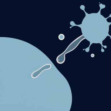
Leukocyte extravasation is a vital process in the body’s immune response, allowing white blood cells (leukocytes) to exit the bloodstream and migrate into tissues where they are needed to combat infections or respond to injury. This complex multistep sequence involves interactions between endothelial cells lining blood vessels and leukocytes circulating in the blood. The efficiency and regulation of this process are essential for maintaining immune surveillance and inflammation control. Any dysregulation in these steps can contribute to a wide range of diseases, including chronic inflammation and autoimmune disorders.
Understanding Leukocyte Extravasation
The Role of Leukocytes
Leukocytes, or white blood cells, play a central role in defending the body against pathogens, removing dead cells, and initiating repair mechanisms. To reach infected or damaged tissues, they must first escape the confines of blood vessels in a tightly regulated manner. This movement is not random but occurs through specific signaling and adhesion mechanisms that guide leukocytes through vessel walls into surrounding tissues.
Importance of the Process
Leukocyte extravasation is crucial during the immune response. It ensures that white blood cells reach sites of injury or infection quickly and efficiently. It also prevents widespread tissue damage by ensuring that leukocytes only enter tissues when necessary. This balance between activation and restraint helps protect the body from both infection and unnecessary inflammation.
Steps of Leukocyte Extravasation
1. Tethering and Rolling
The initial step in leukocyte extravasation is tethering and rolling. In this phase, leukocytes traveling in the bloodstream begin to interact transiently with the endothelial cells of blood vessels. This interaction is mediated by selectins, which are cell adhesion molecules expressed on activated endothelial cells. There are three main types of selectins involved:
- E-selectin: Expressed on endothelial cells during inflammation
- P-selectin: Stored in endothelial cells and platelets, rapidly mobilized
- L-selectin: Found on leukocytes themselves
Selectins bind to specific carbohydrate ligands on leukocytes, allowing the white blood cells to roll along the vessel wall rather than flowing freely. This rolling slows the cells down and brings them into closer contact with the endothelium.
2. Activation
Once leukocytes are rolling along the endothelium, they are exposed to chemokines small signaling proteins secreted by cells at the site of inflammation or injury. These chemokines bind to G protein-coupled receptors on the leukocyte surface, triggering intracellular signaling pathways that lead to the activation of integrins.
This activation is critical because it transforms integrins from a low-affinity to a high-affinity state, enabling the leukocytes to bind more tightly to the endothelial cells. Without chemokine-induced activation, leukocytes would continue rolling without stopping, and the immune response would be inefficient.
3. Firm Adhesion
With integrins now in a high-affinity state, leukocytes can firmly adhere to the endothelium. This step is mediated primarily by interactions between integrins on leukocytes and adhesion molecules on endothelial cells such as:
- ICAM-1(Intercellular Adhesion Molecule-1)
- VCAM-1(Vascular Cell Adhesion Molecule-1)
The most well-known integrins involved include LFA-1 (lymphocyte function-associated antigen-1) and VLA-4 (very late antigen-4). This firm adhesion stops the leukocyte from rolling and anchors it at a specific site along the vessel wall, preparing it for the next phase of extravasation.
4. Transmigration (Diapedesis)
After firm adhesion, leukocytes undergo transmigration, also known as diapedesis. This is the process by which the cell moves between endothelial cells to cross the blood vessel wall. Diapedesis can occur in two ways:
- Paracellular route: Between endothelial cells, which temporarily loosen their junctions
- Transcellular route: Through the body of an individual endothelial cell (less common)
This process involves the leukocyte forming protrusions and squeezing through the endothelial barrier with the help of adhesion molecules like PECAM-1 (Platelet Endothelial Cell Adhesion Molecule-1), JAMs (Junctional Adhesion Molecules), and CD99. Once the leukocyte crosses the endothelial layer, it moves into the subendothelial tissue and continues its journey toward the site of infection or injury.
5. Migration Through the Tissue
After exiting the blood vessel, the leukocyte still needs to travel through the tissue to reach its final destination. This movement is guided by a gradient of chemokines released by damaged cells, pathogens, or resident immune cells. Leukocytes use amoeboid movement to navigate the extracellular matrix, following the chemokine signals. This process ensures that the immune response is localized and efficient.
Molecular Players in Extravasation
Key Adhesion Molecules
Several classes of molecules coordinate the stepwise migration of leukocytes:
- Selectins: Mediate initial rolling
- Integrins: Promote firm adhesion
- Immunoglobulin Superfamily Members: Including ICAM-1 and VCAM-1, aid in firm binding and transmigration
- PECAM-1, CD99, JAMs: Facilitate transmigration
Chemokines and Receptors
Chemokines and their corresponding receptors provide the directional cues that leukocytes follow throughout extravasation. For example:
- CXCL8 (IL-8): Attracts neutrophils
- CCL2: Recruits monocytes
- CCR7: Guides lymphocyte trafficking
Each leukocyte subtype responds to specific chemokines, ensuring that the right type of immune cell reaches the right location.
Physiological and Clinical Relevance
Normal Immune Surveillance
Even in the absence of infection or injury, leukocyte extravasation plays a role in routine immune surveillance. Lymphocytes constantly patrol tissues for signs of abnormal activity, returning to circulation via the lymphatic system if no issues are found.
Inflammatory and Autoimmune Disorders
Dysregulated leukocyte extravasation contributes to numerous health problems. In chronic inflammation, excessive or inappropriate leukocyte infiltration can damage tissues and prolong disease. In autoimmune diseases like rheumatoid arthritis and multiple sclerosis, the immune system attacks healthy tissues, partly due to aberrant leukocyte migration.
Cancer and Tumor Microenvironments
Tumors can exploit the extravasation process by secreting chemokines that attract leukocytes, which may either help or hinder tumor growth. Understanding how leukocytes infiltrate tumors is a key area of research in cancer immunotherapy.
Leukocyte extravasation is a highly coordinated, multistep process that enables white blood cells to travel from the bloodstream to sites of infection, injury, or inflammation. Each stage rolling, activation, adhesion, transmigration, and tissue migration is governed by specific molecular signals and cellular interactions. Proper regulation ensures effective immune defense while minimizing collateral tissue damage. A deeper understanding of this process not only enhances our knowledge of immunology but also opens doors to developing therapies for inflammatory, autoimmune, and cancer-related diseases.