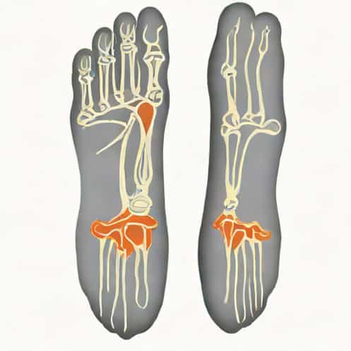
The foot is a complex structure composed of bones, joints, ligaments, muscles, and tendons that work together to support weight, provide balance, and enable mobility. Understanding the anatomy and radiology of the hindfoot, midfoot, and forefoot is crucial for diagnosing injuries, deformities, and degenerative conditions. Radiology provides detailed imaging that allows clinicians to assess bone alignment, joint spaces, fractures, and soft tissue involvement. Proper interpretation of foot radiographs requires knowledge of the normal anatomy and common variations in each region. By examining the hindfoot, midfoot, and forefoot systematically, radiologists and healthcare providers can identify pathologies accurately and provide appropriate treatment recommendations.
Hindfoot Radiology
The hindfoot consists primarily of the talus and calcaneus bones and is responsible for ankle motion, weight transfer, and stability during gait. Radiographic evaluation of the hindfoot is critical for identifying fractures, arthritis, congenital deformities, and post-traumatic changes. Standard imaging includes anteroposterior, lateral, and oblique radiographs, which help visualize the alignment and morphology of the bones and joints.
Anatomy and Key Landmarks
Key landmarks in the hindfoot include the talar dome, calcaneal tuberosity, subtalar joint, and sinus tarsi. The talus articulates with the tibia and fibula to form the ankle joint and with the calcaneus to form the subtalar joint, enabling inversion and eversion movements. The calcaneus bears the majority of weight and serves as the attachment site for the Achilles tendon. Accurate identification of these structures on radiographs is essential for detecting fractures, dislocations, or degenerative changes.
Common Hindfoot Pathologies
- Calcaneal fractures often due to high-impact trauma; radiographs help assess displacement and joint involvement.
- Talus fractures particularly the talar neck, which may compromise vascular supply and lead to avascular necrosis.
- Subtalar arthritis degenerative changes visible as joint space narrowing and osteophyte formation.
- Hindfoot valgus or varus deformities alignment assessment is essential for surgical planning.
Midfoot Radiology
The midfoot comprises the navicular, cuboid, and cuneiform bones, forming the arches of the foot that provide stability and shock absorption. Midfoot radiology focuses on detecting fractures, dislocations, arthritis, and ligamentous injuries. Imaging typically involves weight-bearing anteroposterior and lateral views, which are crucial for evaluating arch height and alignment.
Anatomy and Key Landmarks
Important midfoot structures include the tarsometatarsal joints (Lisfranc joints), the navicular tuberosity, and the cuboid articulation with the calcaneus and metatarsals. The stability of the midfoot is largely dependent on the integrity of ligaments, particularly the Lisfranc ligament, which connects the medial cuneiform to the second metatarsal. Accurate radiographic assessment helps identify subtle fractures, dislocations, or joint subluxations that may compromise foot mechanics.
Common Midfoot Pathologies
- Lisfranc injuries range from ligamentous tears to fracture-dislocations; weight-bearing radiographs are essential for detection.
- Navicular stress fractures common in athletes and may present as subtle radiographic changes.
- Osteoarthritis degenerative changes can affect tarsometatarsal joints, visible as joint space narrowing and sclerosis.
- Midfoot collapse often secondary to neuropathic conditions or trauma, visible as loss of the longitudinal arch.
Forefoot Radiology
The forefoot includes the metatarsals, phalanges, and associated joints and soft tissues. It is responsible for weight distribution during propulsion, balance, and fine movements of the toes. Radiographic assessment of the forefoot is important for evaluating fractures, deformities such as bunions and hammer toes, and degenerative or inflammatory joint disease. Standard imaging includes anteroposterior, lateral, and oblique views.
Anatomy and Key Landmarks
Key forefoot structures include the metatarsophalangeal joints, interphalangeal joints, and sesamoid bones beneath the first metatarsal head. These structures facilitate toe motion and absorb forces during walking and running. Understanding the normal relationships of the metatarsals, the alignment of the phalanges, and the position of the sesamoids is critical for identifying fractures, dislocations, and alignment abnormalities.
Common Forefoot Pathologies
- Metatarsal fractures common in sports injuries and falls; radiographs help determine displacement and healing progress.
- Phalangeal fractures may involve distal, middle, or proximal phalanges, with or without joint involvement.
- Hallux valgus and bunion deformities lateral deviation of the great toe, assessable through AP and oblique views.
- Osteoarthritis and gout degenerative or inflammatory changes are visible as joint space narrowing, erosions, or osteophyte formation.
- Sesamoid fractures often occur due to repetitive stress and are best seen in specialized axial views.
Imaging Techniques and Considerations
Radiography remains the primary modality for evaluating the hindfoot, midfoot, and forefoot, but advanced imaging techniques such as computed tomography (CT) and magnetic resonance imaging (MRI) are increasingly used for complex cases. CT provides detailed bone imaging, ideal for subtle fractures and preoperative planning, while MRI excels in assessing soft tissue, ligaments, and bone marrow changes. Weight-bearing radiographs are particularly important for assessing alignment and functional deformities, providing insight into the biomechanical consequences of injuries or chronic conditions.
Tips for Radiographic Interpretation
- Always evaluate alignment and joint spaces across all foot regions.
- Compare both feet when possible to detect subtle differences.
- Identify normal anatomic variants to avoid misdiagnosis.
- Use oblique and specialized views for sesamoids, midfoot, and complex fractures.
- Correlate clinical findings with imaging to guide treatment decisions.
Radiologic assessment of the hindfoot, midfoot, and forefoot is a cornerstone of foot care, enabling accurate diagnosis of fractures, dislocations, deformities, and degenerative conditions. Understanding the anatomy, key landmarks, and common pathologies of each region is essential for proper evaluation. Weight-bearing radiographs, CT, and MRI complement each other to provide a comprehensive view of the foot’s structure and function. By systematically examining each segment and interpreting imaging findings in the context of clinical presentation, radiologists and clinicians can ensure effective management, improved patient outcomes, and preservation of foot health. Mastery of hindfoot, midfoot, and forefoot radiology is therefore critical for healthcare providers involved in orthopedics, sports medicine, and podiatry.