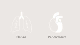
The human body is protected and supported by various membranes that surround vital organs, ensuring both structure and function. Two of the most important membranes in the thoracic cavity are the pleura and the pericardium. These specialized serous membranes are responsible for enclosing the lungs and the heart, respectively. While each has its own unique anatomical location and purpose, they share a common structural composition and function in reducing friction, protecting internal organs, and facilitating smooth movement during breathing and cardiac activity. Understanding what makes up the pleura and pericardium provides key insight into thoracic anatomy and physiology.
Overview of Serous Membranes
Both the pleura and pericardium are examples ofserous membranes, also known as serosa. These membranes line internal cavities and cover organs within them. They are composed of two main layers:
- Parietal layer– Lines the internal surface of the body cavity wall.
- Visceral layer– Covers the surface of the organ itself.
The space between these layers is filled with a small amount of serous fluid, which acts as a lubricant to prevent friction as the organs move.
Tissue Composition of Pleura and Pericardium
The pleura and pericardium are both primarily made up of a combination ofmesotheliumandconnective tissue. These layers are adapted to their protective and lubricating functions.
Mesothelial Cells
Mesothelium is a single layer of flattened squamous epithelial cells. These cells are specialized for secretion and absorption of serous fluid. They provide a smooth, slippery, and non-adhesive surface that reduces friction between moving organs.
Connective Tissue Layer
Beneath the mesothelium is a thin layer of loose connective tissue. This layer contains collagen fibers, elastic fibers, blood vessels, lymphatics, and nerves. It provides structural support and elasticity to the membrane and helps anchor it in place.
The Pleura: Structure and Function
The pleura surrounds the lungs and lines the thoracic cavity. Each lung is enclosed in its own pleural sac. The pleura consists of two main parts:
- Parietal pleura– Lines the thoracic wall, diaphragm, and mediastinum.
- Visceral pleura– Closely adheres to the surface of the lungs.
Pleural Cavity
Between the parietal and visceral pleura is the pleural cavity, which contains pleural fluid. This fluid allows the lungs to expand and contract smoothly during respiration without causing friction against the thoracic wall.
Functions of the Pleura
- Reduces friction between lung tissue and chest wall during breathing.
- Creates surface tension that helps keep the lungs inflated.
- Serves as a barrier to infection and fluid leakage.
The Pericardium: Structure and Function
The pericardium surrounds the heart and roots of the great vessels. It also has two layers, similar in structure to the pleura:
- Fibrous pericardium– A tough outer layer made of dense connective tissue.
- Serous pericardium– Subdivided into the parietal and visceral pericardium (also called the epicardium).
Pericardial Cavity
The pericardial cavity is the space between the parietal and visceral layers of the serous pericardium. It contains a small amount of pericardial fluid that reduces friction as the heart beats and moves within the thoracic cavity.
Functions of the Pericardium
- Anchors the heart in position within the mediastinum.
- Prevents excessive movement or overexpansion of the heart.
- Provides a frictionless environment for cardiac function.
- Acts as a physical barrier against infection or inflammation from adjacent organs.
Differences Between Pleura and Pericardium
While both are serous membranes, the pleura and pericardium differ in terms of their location, structure, and specific function.
Location
- Pleura– Associated with the lungs and thoracic cavity.
- Pericardium– Encloses the heart and the bases of the great vessels.
Structure
- Pleura– Has only a serous layer (parietal and visceral pleura).
- Pericardium– Has both fibrous and serous layers.
Fluid Contents
- Pleural fluid– Approximately 10-20 ml in healthy individuals.
- Pericardial fluid– Normally about 15-50 ml in adults.
Clinical Relevance of Pleura and Pericardium
Because of their protective and supportive roles, disorders affecting these membranes can have serious health implications.
Pleural Disorders
- Pleuritis (Pleurisy)– Inflammation of the pleura causing sharp chest pain during breathing.
- Pleural effusion– Accumulation of excess fluid in the pleural cavity.
- Pneumothorax– Air in the pleural space leading to lung collapse.
Pericardial Disorders
- Pericarditis– Inflammation of the pericardium, often causing chest pain and friction rub.
- Pericardial effusion– Excess fluid buildup that may compress the heart.
- Cardiac tamponade– Life-threatening condition caused by fluid pressure impairing heart function.
Embryological Origin
Both the pleura and pericardium develop from the embryonic mesoderm, specifically the lateral plate mesoderm. The serous membranes form from the splanchnic and somatic layers of the mesoderm:
- Visceral layers– Derived from splanchnic mesoderm, associated with the organs.
- Parietal layers– Derived from somatic mesoderm, associated with body wall structures.
This shared origin explains their similar histological structure and functional roles.
Microscopic Anatomy
Under the microscope, both membranes show the following layers:
- Mesothelial lining– Simple squamous epithelium, smooth and frictionless.
- Basement membrane– Anchors the mesothelium to underlying tissues.
- Loose connective tissue– Contains capillaries, lymphatics, and nerves for support and communication.
This arrangement allows rapid diffusion of gases and efficient fluid production and absorption.
The pleura and pericardium are essential components of the thoracic cavity, composed of mesothelium and connective tissue. These serous membranes protect the lungs and heart, reduce friction, and support the physiological movements of breathing and circulation. Though they serve different organs, their structural similarities highlight the body’s efficient design in maintaining function and minimizing injury. Understanding what makes up the pleura and pericardium is not only important in anatomical studies but also in diagnosing and managing conditions that affect respiratory and cardiovascular health.