The normal cortical thickness of the kidney is a key parameter in assessing renal health and function. The renal cortex is the outer layer of the kidney, housing essential structures such as glomeruli and convoluted tubules, which play a critical role in filtration and urine formation. Evaluating the thickness of the renal cortex is important in diagnosing and monitoring kidney diseases, including chronic kidney disease, renal atrophy, and other structural abnormalities. Imaging techniques, particularly ultrasound and CT scans, are widely used to measure cortical thickness, providing valuable insights into kidney morphology, functionality, and overall health.
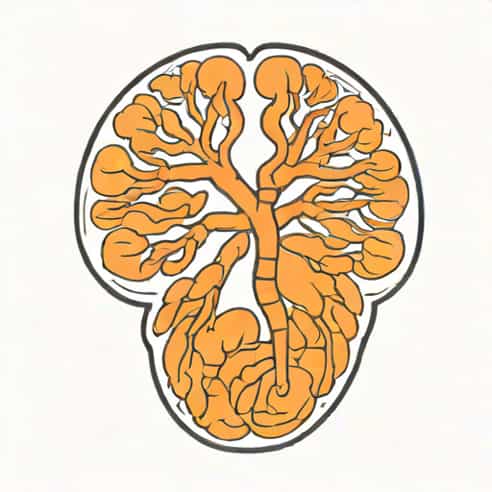
Anatomy of the Renal Cortex
The renal cortex forms the outermost layer of the kidney, lying beneath the fibrous capsule and surrounding the medulla. It contains vital components like glomeruli, proximal and distal convoluted tubules, and interstitial tissue, all of which are integral to kidney function. The cortex receives a significant portion of renal blood flow, highlighting its importance in filtration and metabolic regulation. Understanding the normal cortical thickness is essential for distinguishing between healthy and pathological kidneys, as changes in thickness often correlate with renal dysfunction.
Normal Cortical Thickness Values
Normal cortical thickness can vary depending on age, body size, and overall health. In adults, the renal cortex typically measures between 7 mm and 10 mm on imaging studies such as ultrasound. Pediatric populations have thinner cortices, reflecting smaller kidney size and developmental stages. Maintaining awareness of these normal ranges allows clinicians to identify deviations that may indicate disease or chronic damage. Measurement should be performed at the mid-pole of the kidney, avoiding regions where the cortex may be naturally thinner due to vascular structures or the renal hilum.
- Adult normal cortical thickness approximately 7-10 mm
- Variations based on age and kidney size
- Measurement location mid-pole of the kidney for accuracy
- Importance in assessing renal function and disease progression
Factors Affecting Cortical Thickness
Cortical thickness may be influenced by multiple physiological and pathological factors. Age-related cortical thinning is a natural process, often accompanied by decreased renal function. Conditions such as chronic kidney disease, hypertension, diabetes mellitus, and glomerulonephritis can also lead to significant cortical thinning. Conversely, cortical thickening may occur in certain conditions, such as acute kidney injury or compensatory hypertrophy following unilateral nephrectomy. Understanding these factors is crucial for proper interpretation of imaging findings and appropriate clinical decision-making.
Age and Development
During childhood, the renal cortex gradually thickens as the kidneys grow. Peak cortical thickness is typically observed in early adulthood, after which a gradual decline occurs due to age-related nephron loss and vascular changes. Monitoring cortical thickness over time can help identify early signs of renal aging or pathology before clinical symptoms manifest.
Renal Pathologies
Various renal pathologies can significantly alter cortical thickness
- Chronic kidney disease (CKD)Progressive nephron loss leads to cortical thinning, often associated with decreased kidney size and altered echogenicity.
- Diabetic nephropathyChronic hyperglycemia causes structural damage, potentially resulting in cortical thinning and increased echogenicity on ultrasound.
- GlomerulonephritisInflammatory processes may cause early cortical swelling, followed by chronic thinning if untreated.
- Acute kidney injuryCortical thickening may be observed due to edema and inflammation.
Imaging Techniques for Measuring Cortical Thickness
Accurate measurement of cortical thickness is primarily achieved through imaging modalities. Ultrasound is the most commonly used method due to its non-invasive nature, real-time visualization, and cost-effectiveness. CT and MRI provide additional detail, particularly in complex cases where precise anatomical mapping is necessary. Each modality has specific advantages and considerations in assessing the renal cortex.
Ultrasound Measurement
Ultrasound evaluation involves placing the transducer along the kidney’s longitudinal axis to measure cortical thickness at the mid-pole. The cortex appears as a hypoechoic layer between the echogenic renal capsule and the brighter medulla. Consistent measurement techniques are crucial to reduce inter-observer variability and ensure reliable data for clinical assessment.
- Non-invasive and widely accessible
- Real-time visualization of cortical thickness and kidney morphology
- Operator-dependent accuracy; requires standardized protocols
CT and MRI Assessment
CT imaging allows high-resolution measurement of cortical thickness, particularly when evaluating complex anatomical or pathological conditions. MRI provides superior soft tissue contrast, making it suitable for detailed assessment of renal parenchyma and early detection of cortical changes. Both modalities are valuable adjuncts to ultrasound in comprehensive kidney evaluation, especially in patients with obesity, poor acoustic windows, or complex renal disease.
Clinical Significance of Cortical Thickness
Evaluating cortical thickness is essential in diagnosing, monitoring, and managing kidney diseases. Thinning of the cortex often correlates with reduced glomerular filtration rate (GFR) and impaired kidney function, serving as an early marker for chronic kidney disease. Conversely, abnormal thickening may indicate acute injury, inflammation, or compensatory hypertrophy. Tracking changes over time helps clinicians assess disease progression, guide treatment, and predict patient outcomes.
Applications in Kidney Disease Monitoring
- Early detection of chronic kidney disease through cortical thinning assessment
- Monitoring disease progression in diabetic nephropathy and glomerulonephritis
- Evaluating response to therapies such as antihypertensive or immunosuppressive treatments
- Predicting renal function decline and need for interventions
The normal cortical thickness of the kidney is a vital parameter in assessing renal health. Understanding the typical ranges, factors affecting cortical thickness, and imaging techniques for measurement allows clinicians to detect and monitor kidney diseases effectively. Regular evaluation of cortical thickness, combined with other clinical and laboratory parameters, provides comprehensive insights into kidney function, disease progression, and patient prognosis. By maintaining awareness of normal cortical dimensions and interpreting changes accurately, healthcare providers can optimize care, prevent complications, and ensure better outcomes for patients with renal conditions.
In summary, the renal cortex is essential for filtration and overall kidney function, and its thickness is a valuable indicator of health. Accurate measurement using ultrasound, CT, or MRI, alongside understanding the effects of age, disease, and other factors, allows clinicians to make informed decisions. Continuous monitoring of cortical thickness can serve as a predictive and diagnostic tool, ensuring that individuals receive timely interventions and appropriate management for kidney-related health issues. Awareness and evaluation of normal cortical thickness remain fundamental to nephrology and general medical practice.