Proper X-ray angulation for maxillary molars is a critical aspect of dental radiography, ensuring accurate diagnosis, treatment planning, and successful endodontic or restorative procedures. Maxillary molars, due to their complex root anatomy and position in the posterior maxilla, require precise angulation to capture all root canals, periapical structures, and surrounding bone clearly. Understanding the principles of X-ray beam angulation, the use of different radiographic techniques, and common challenges associated with maxillary molar imaging is essential for dental professionals to achieve high-quality diagnostic images and improve patient outcomes.
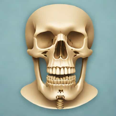
Anatomy of Maxillary Molars and Radiographic Considerations
Maxillary molars typically have three roots the mesiobuccal, distobuccal, and palatal roots, with the mesiobuccal root often containing two canals. The roots are widely divergent, especially the palatal root, which can pose challenges in achieving clear radiographic images. Additionally, maxillary molars are located near the maxillary sinus, which can obscure root apices if angulation is incorrect. Understanding the anatomical variations and surrounding structures is crucial for selecting the correct X-ray angulation.
Principles of X-Ray Angulation
X-ray angulation involves the orientation of the X-ray beam in relation to the tooth and the image receptor. Proper angulation minimizes distortion, superimposition of anatomical structures, and ensures that the image accurately represents the true anatomy of the maxillary molars. There are two primary aspects of angulation to consider
- Horizontal AngulationRefers to the side-to-side orientation of the X-ray beam. Incorrect horizontal angulation can result in overlapping roots or missed canals.
- Vertical AngulationRefers to the up-and-down tilt of the X-ray beam. Proper vertical angulation ensures that the roots are captured without foreshortening or elongation.
Periapical Radiography for Maxillary Molars
Periapical radiographs are the most common method for evaluating maxillary molars. The paralleling technique is preferred over the bisecting-angle technique due to its ability to reduce image distortion and provide more consistent results. In the paralleling technique, the image receptor is placed parallel to the long axis of the tooth, and the X-ray beam is directed perpendicular to both the tooth and the receptor.
Vertical Angulation Guidelines
For maxillary molars, a slight positive vertical angulation is typically recommended. This means that the X-ray tube is angled slightly downward relative to the horizontal plane. Generally, a vertical angulation of approximately +20 to +25 degrees is used to compensate for the upward tilt of the maxillary arch. This adjustment helps to project the roots clearly onto the image receptor, minimizing overlap and distortion.
Horizontal Angulation Guidelines
Horizontal angulation is equally important to avoid superimposition of the roots. For maxillary molars, the X-ray beam should be directed perpendicular to the mesiodistal plane of the tooth. Slight mesial or distal shifts can be applied to visualize individual canals within multirooted teeth, particularly when performing endodontic procedures. Proper horizontal angulation ensures that the mesiobuccal, distobuccal, and palatal roots are visible as separate entities on the radiograph.
Advanced Radiographic Techniques
In addition to traditional periapical radiographs, advanced imaging modalities such as cone beam computed tomography (CBCT) can provide three-dimensional visualization of maxillary molars. CBCT eliminates many of the limitations of two-dimensional imaging by allowing dental professionals to examine root morphology, canal configuration, and periapical structures from multiple angles. While CBCT is not always necessary for routine evaluations, it is particularly useful in complex cases or when conventional radiographs are inconclusive.
Challenges in Maxillary Molar Imaging
Several factors can complicate the radiographic imaging of maxillary molars
- Sinus SuperimpositionThe maxillary sinus can obscure root apices, particularly the palatal root.
- Patient AnatomyHigh palatal vaults or limited mouth opening can make proper receptor placement difficult.
- Root MorphologyDivergent or curved roots can require multiple angulations to visualize all canals.
- Artifacts and MovementPatient movement or improper receptor positioning can lead to blurred or distorted images.
Clinical Tips for Optimal Angulation
To achieve high-quality radiographs of maxillary molars, dental professionals should follow these tips
- Use the paralleling technique whenever possible to minimize distortion.
- Adjust vertical angulation to approximately +20 to +25 degrees for maxillary molars.
- Ensure horizontal angulation is perpendicular to the mesiodistal plane of the tooth, adjusting slightly to separate overlapping roots.
- Take multiple radiographs from different angles if necessary, particularly for multirooted teeth.
- Consider CBCT for complex cases, including retreatment or assessment of anatomical variations.
- Ensure patient comfort and proper receptor placement to reduce movement and enhance image clarity.
Importance in Endodontic and Restorative Procedures
Proper X-ray angulation for maxillary molars is crucial in endodontic treatments, as it allows clinicians to identify all root canals, assess curvature, and detect periapical pathology. In restorative dentistry, accurate radiographs inform decisions regarding crowns, bridges, and implants. Misaligned angulation can result in missed canals, incorrect diagnosis, or procedural complications, underscoring the need for precise technique and attention to detail.
X-ray angulation for maxillary molars is a fundamental skill in dental radiography that directly impacts diagnostic accuracy and treatment outcomes. By understanding the anatomical complexities of maxillary molars, applying proper vertical and horizontal angulation, and utilizing advanced imaging techniques when necessary, dental professionals can obtain high-quality radiographs that guide effective clinical decisions. Attention to patient positioning, careful technique, and adherence to radiographic principles ensures that maxillary molars are accurately visualized, facilitating successful endodontic, restorative, and surgical interventions. Mastery of X-ray angulation for maxillary molars is essential for delivering precise, safe, and effective dental care.